Description
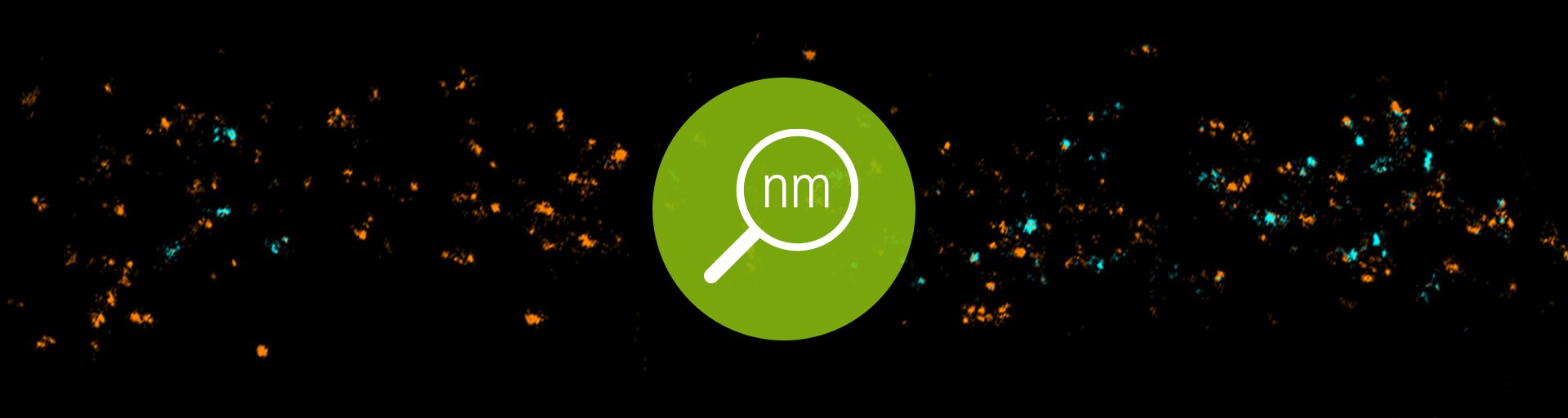
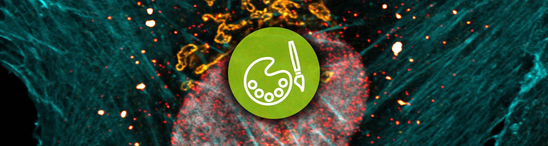
abberior STAR ORANGE is a novel fluorescent dye developed for STED and confocal microscopy in the orange spectral region. Introducing negatively charged groups into its molecular structure making the dye excellently water soluble and enables background-free imaging. Thus, abberior STAR ORANGE is highly photostable and a bright orange fluorescent dye, which can be effectively excited with excitation light between 550 - 610 nm. For STED microscopy, this dye can be most efficiently used with a STED laser wavelength between 750 - 800 nm. Our abberior STAR ORANGE can substitute dyes like ATTO® 594 or Alexa Fluor® 594. Together with our abberior STAR RED you can obtain stunning 2 color STED results. In combination with our abberior STAR 580 you get the ideal pair for FLIM experiments. Best results are obtained with freshly prepared samples.
NEW: Learn more about our latest variants conjugated to Smart Secondaries® (nanobodies) optimized for efficient labeling and minimal linkage error in STED and confocal microscopy.
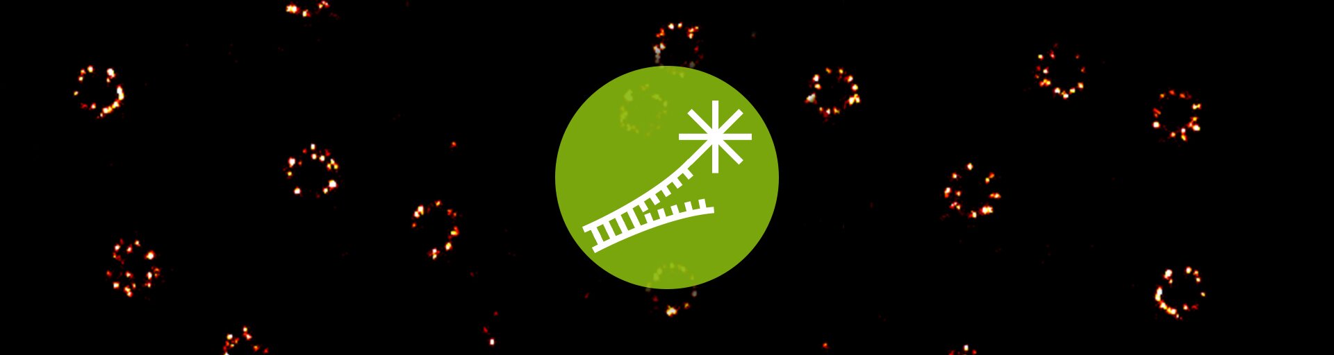
Secondary single-domain antibodies (sdABs), also known as nanobodies, represent an innovative and promising antibody format. At just 15 kDa, sdABs are 10 times smaller than conventional whole IgG antibodies, making them ideal for high-resolution imaging applications that require small labels, excellent tissue penetration, and exceptional specificity. The combination of abberior STAR dyes – known for their superior brightness and photostability – with secondary sdABs (FluoTag®-X2) from NanoTag Biotechnologies provides an exceptional solution for high-quality, high-resolution imaging.
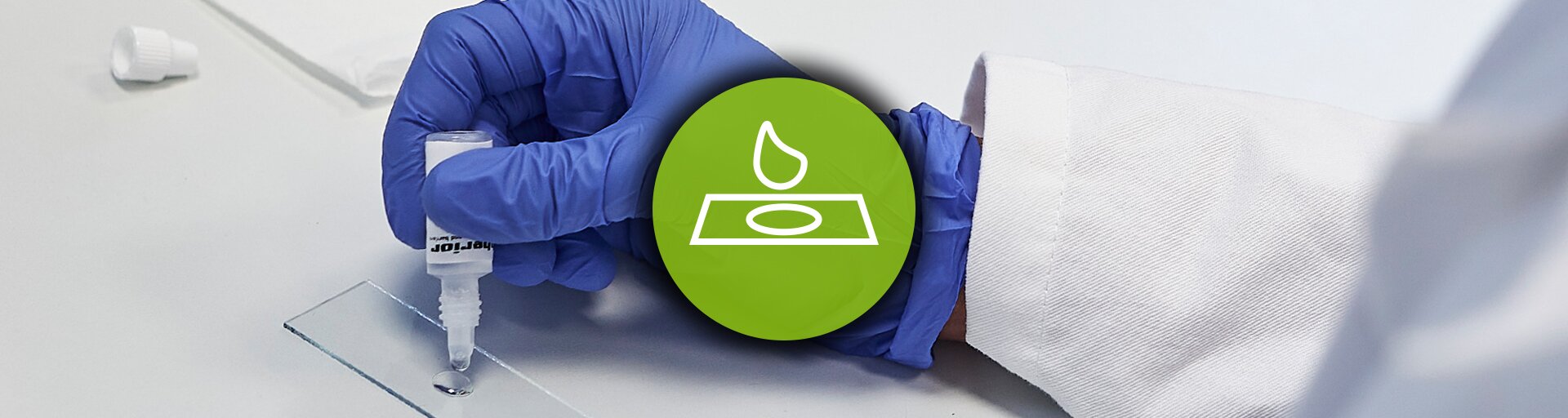
FluoTag®-X2 sdABs, developed by NanoTag Biotechnologies, bind with high affinity and specificity to a distinct epitope on mouse IgG1. Due to the inherent symmetry of the target antibody, two secondary nanobodies will be able to bind each primary antibody. The secondary FluoTag®-X2 reagents will therefore target 4 fluorophores to each primary antibody. This product includes one abberior STAR ORANGE FluoTag®-X2 anti-Mouse IgG1 vial as a lyophilized reagent. Before staining, dissolve the reagent in 500 µl of sterile 50% glycerol (v/v) in deionized water to obtain a concentration of 5 µM of the nanobody (10 µM dye). Please refer to our protocol for detailed instructions on the handling, storage, and staining procedure.